渋谷TMJ歯科クリニック|頭蓋・頸椎と顎位・咬合の関係について
渋谷TMJ歯科クリニックでは、頭蓋および頸椎の歪みが顎位(あごの位置)と咬合(噛み合わせ)の乱れを引き起こし、その結果として顔の歪み、顎関節症、食いしばり、ストレートネックによる強い肩こり、さらには自律神経の乱れへとつながると考えています。
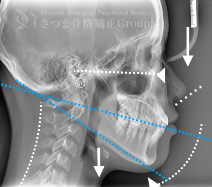
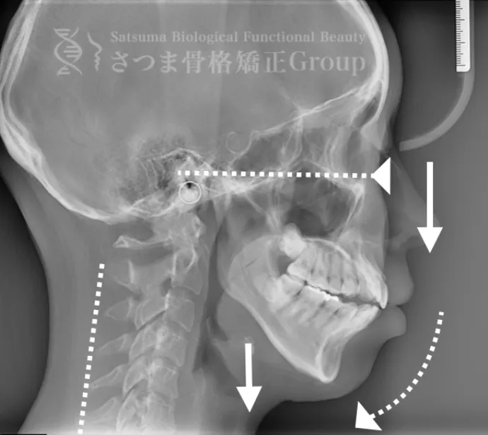
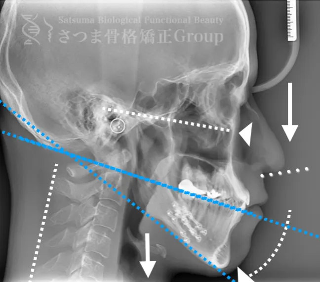
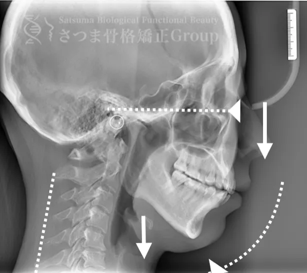
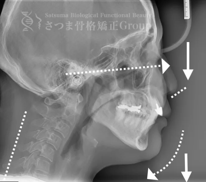
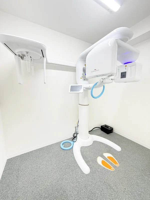
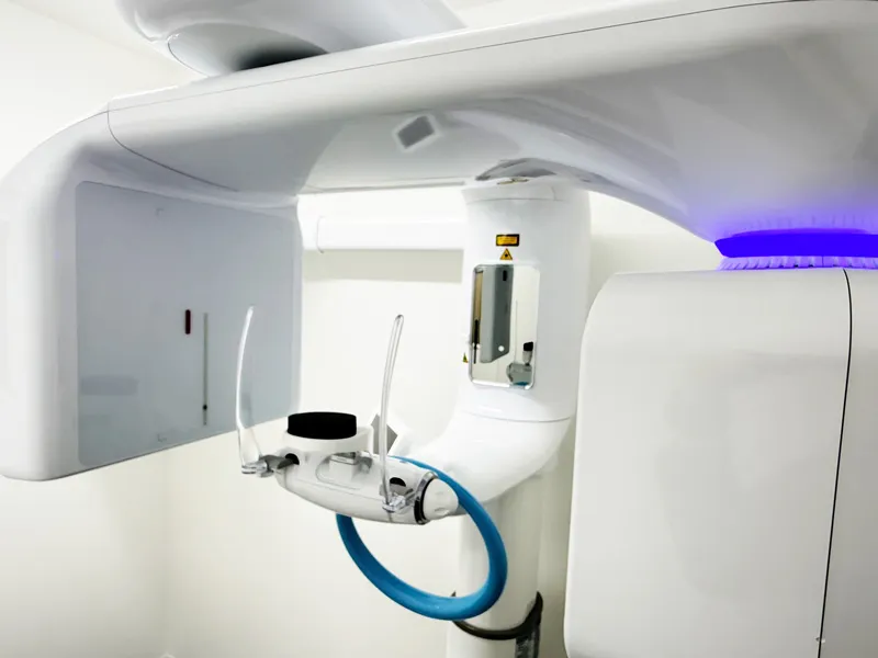
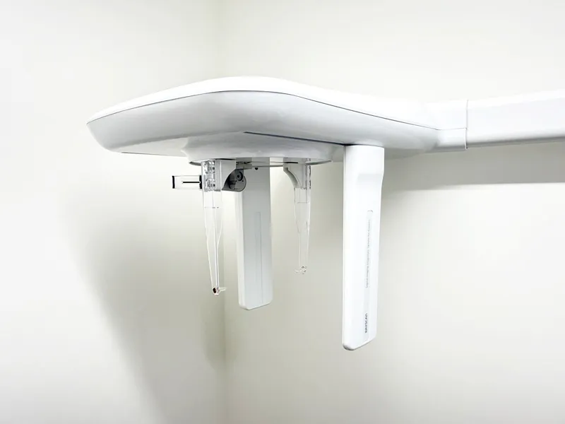
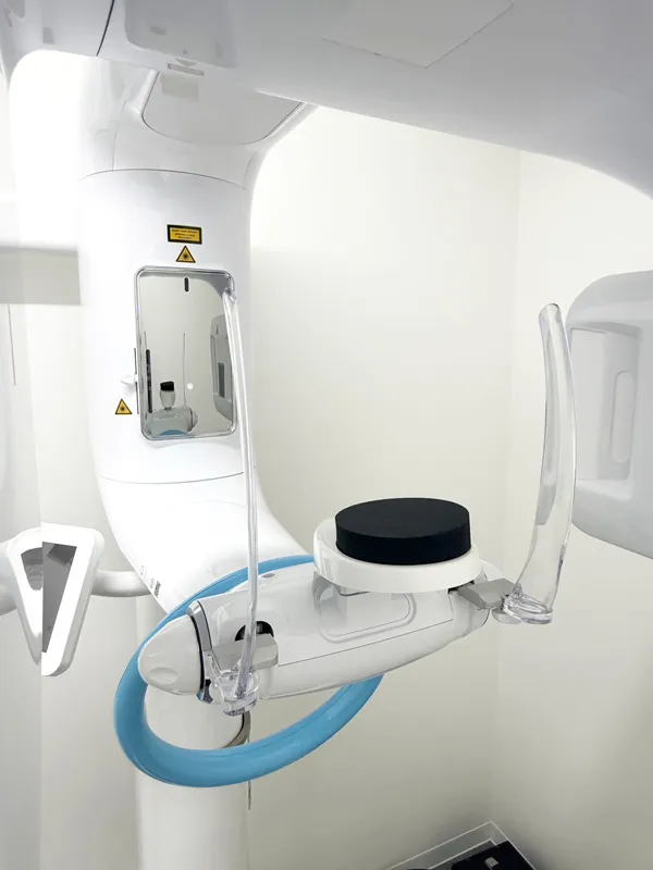
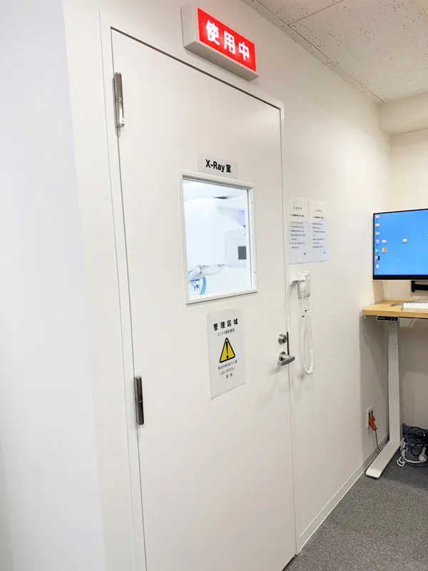
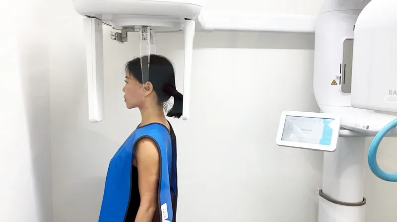
これらの問題を正確に評価するため、当院では セファロシステムによるレントゲン撮影(X-ray images) を行っています。
セファロ撮影によって 顎関節・頭蓋骨・頸椎の位置関係を客観的に可視化でき、施術前の状態を科学的に把握することが可能となります。
その結果、
• 顎位のズレ
• 咬合の乱れ
• 頭位・頸椎姿勢のバランス
• 筋肉・関節の負担部位
を精密に分析し、
患者様ごとに最適な施術プランを立て、歯科治療+骨格施術をしていきます。

