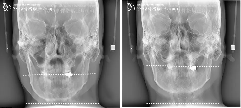
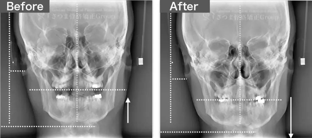
The mandible shows a slight leftward shift with instability (slippage) of the mandibular position.
AFTER
中顔面の短縮 下顎の左方シフト改善、 正中誘導 下顎位の改善 整顔率の向上
correction of the leftward mandibular shift; guidance toward the facial midline; improvement in mandibular positioning; increased facial balance and symmetry.
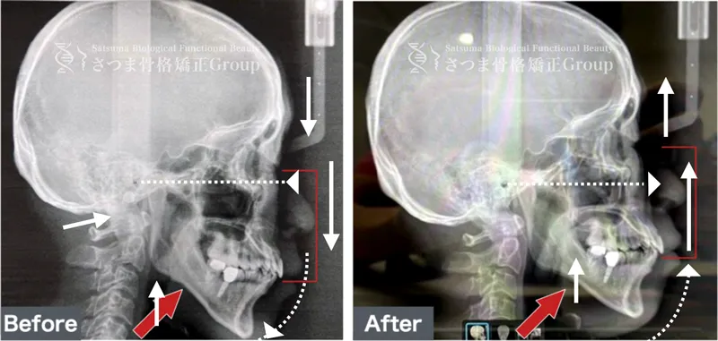
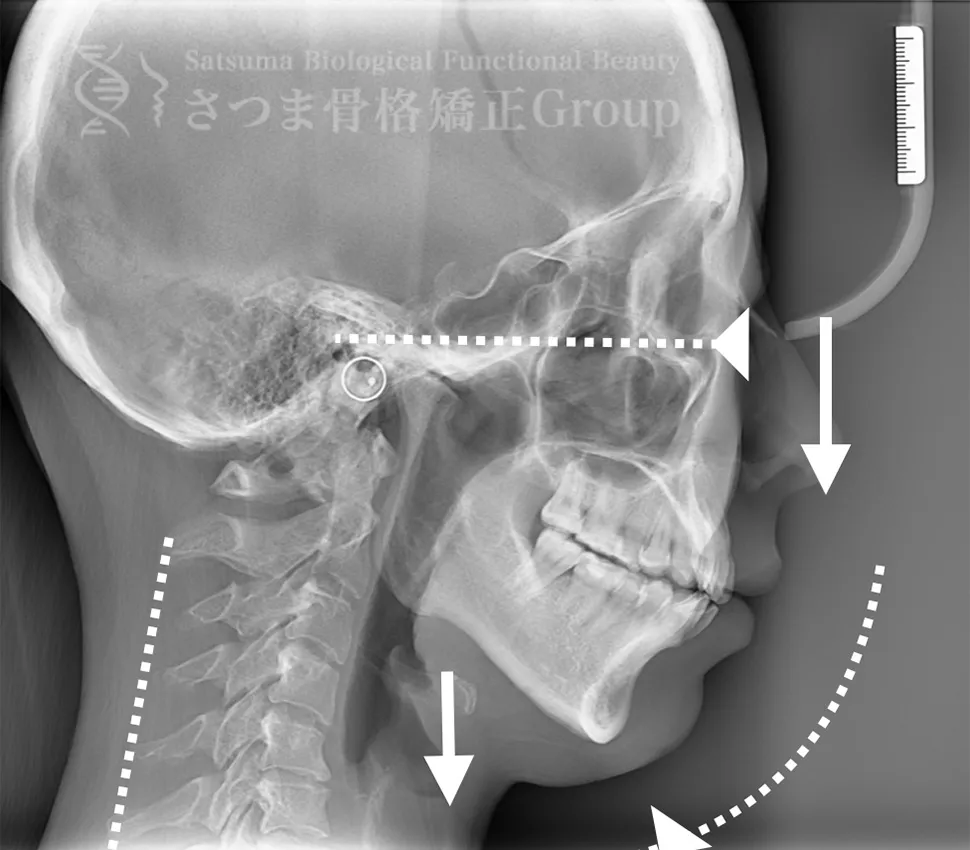
Mild jaw deformity present. Posterior rotation of the mandible. The midface is elongated and has dropped downward. Loss of depth in the midface. Cervical spine straightening (straight neck).
Release the masseter and upper cervical spine to achieve forward rotation of the jaw.
Using the Satsuma method, create depth in the midface and shorten it.
Enhance the cervical lordosis angle.
Posterior rotation of the mandible The midface is elongated and has dropped downward Loss of depth in the midface Cervical spine straightening (straight neck) Severe neck stiffness
After the osteopathy, the custom-formed mouthpiece guides the shape and promotes forward rotation of the temporomandibular joint. It facilitates the release of the upper cervical spine and encourages the body’s natural self-correcting ability. Mobilization of the sphenopalatine suture and the sphenomaxillary suture boosts mobility and shortens the midface.
BEFORE
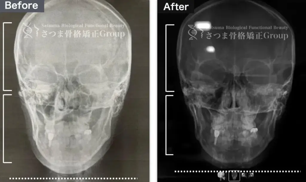
口蓋骨の下方変位 下顎位の滑落 ストレートネック 中顔面と人中が伸びて見える
Inferior displacement of the palatine bone Sagging of the mandibular position Straight neck The midface and philtrum appear elongated
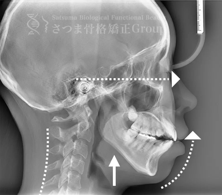
AFTER
口蓋骨を上方に整復 下顎位が上がる ストレートネックの改善 中顔面と人中が短縮し小顔に見える
Repositioning the palatine bone upward The mandibular position is elevated Improvement of straight neck The midface and philtrum are shortened, giving the appearance of a smaller face
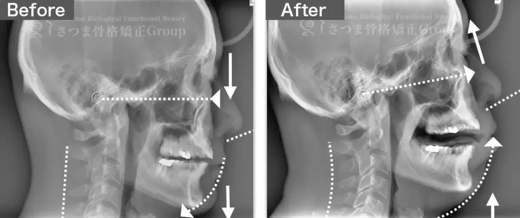
20代女性 S様 初回効果
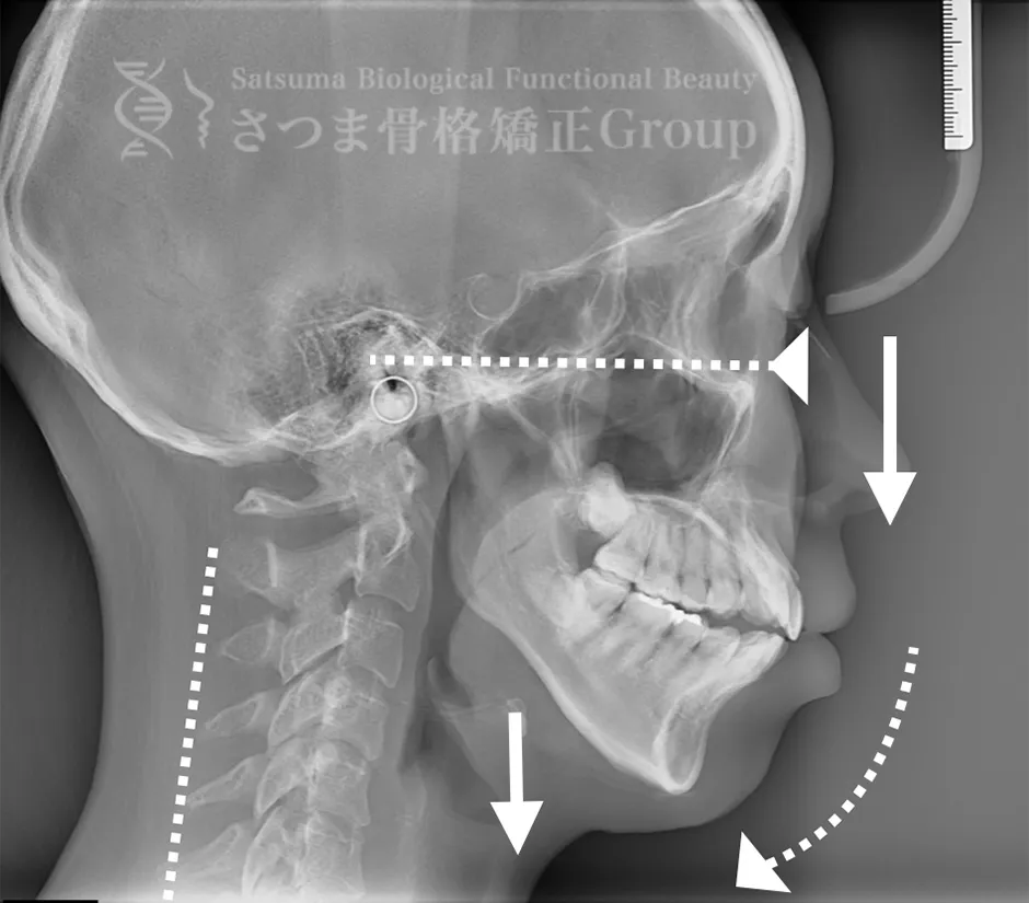
BEFORE
下顎の後方回転あり
中顔面が伸びて下方に落ちる
中顔面の奥行きを失っている
頚椎ストレートネック
Posterior rotation of the mandible, elongated and downward-dropped midface, loss of midface depth, and cervical spine straightening (straight neck).
Release the masseter and upper cervical spine to achieve forward rotation of the jaw. Using the Satsuma method, create depth in the midface and shorten it. Enhance the cervical lordosis angle.
After the osteopathy, maintain the shape and the release of the upper cervical spine with a custom-formed mouthpiece, and boost the mobility of the sphenoid bone while aligning the facial bones for facial correction and structural adjustment.
Mild jaw deformity present. Posterior rotation of the mandible. The midface is elongated and has dropped downward. Loss of depth in the midface. Cervical spine straightening (straight neck).
Release the masseter and upper cervical spine to achieve forward rotation of the jaw, use the Satsuma method to create and shorten midface depth, and enhance the cervical lordosis angle.
After the osteopathy, the custom-formed mouthpiece maintains the shape and the release of the upper cervical spine, boosts the mobility of the sphenoid bone, and aligns the facial bones for facial correction and structural adjustment.
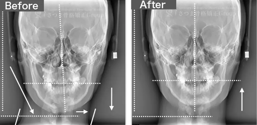
BEFORE
軽度の顎変形症あり 下顎の左方シフト 頚椎の斜頸 中顔面の滑落
Mild jaw deformity present Leftward shift of the mandible Cervical spine torticollis Sagging of the midface
AFTER
下顎の正中誘導 頚椎の斜頸矯正 中顔面の短縮
Midline correction of the mandible Correction of cervical spine torticollis Shortening of the midface